


Erler-Zimmer Sagittal head section of a horse

SKU: SKU: EZVP10000
PZN: PZN:
Packaging unit: VE:
Inbound: deliverable probably in 2-3 weeks
Verpackungseinheit :
Couldn't load pickup availability


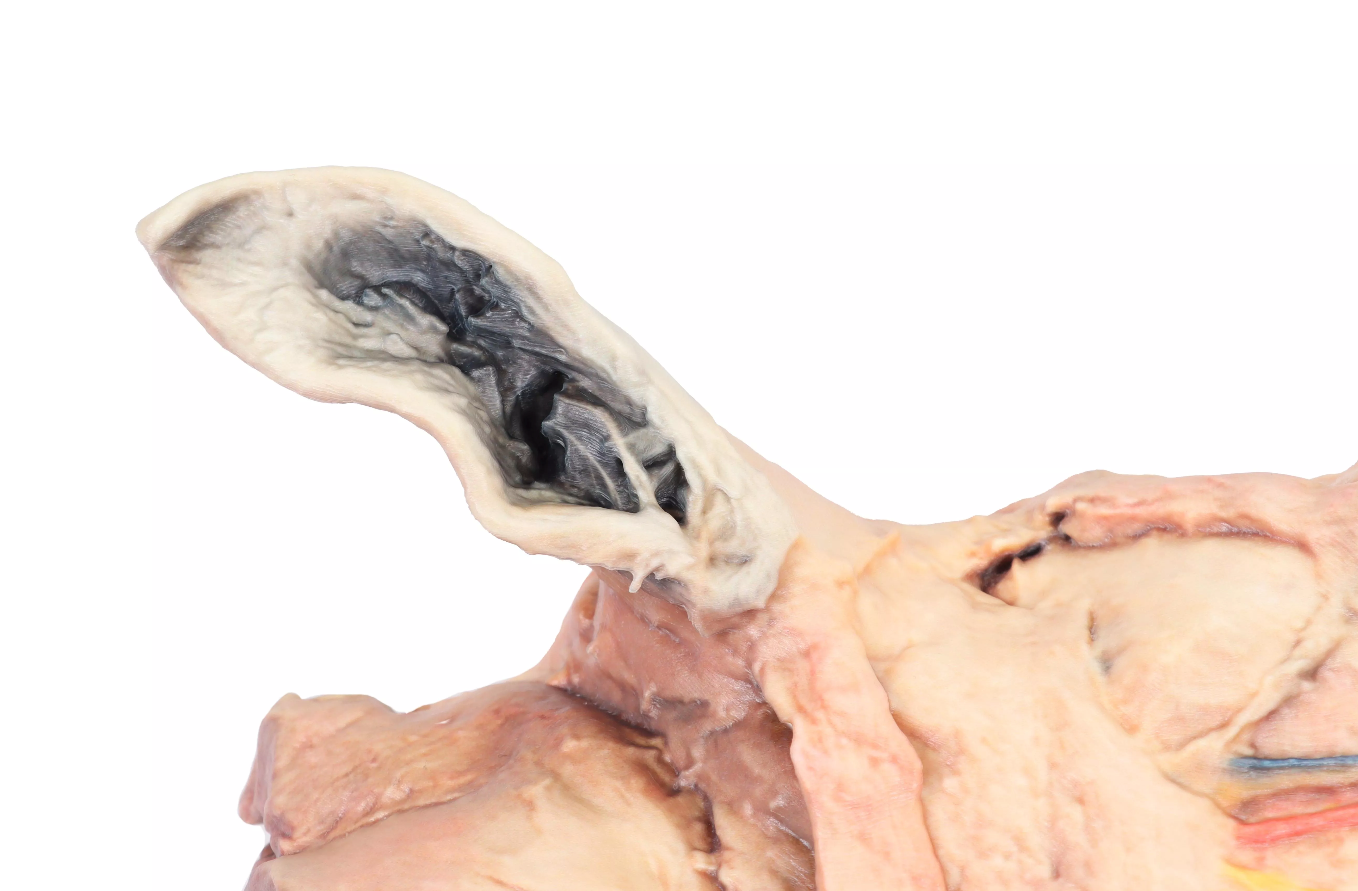
This high-quality anatomical model of a half horse head is an indispensable teaching tool for veterinary education. It offers a dual representation: The right lateral side shows a precise superficial dissection of the musculature and nerve pathways, while the medial median sagittal section provides deeper insights into the respiratory tract, digestive system, and central nervous system.
Lateral View (Surface Anatomy)
On the exterior, the skin has been removed (except for nostrils, lips, and auricle) to reveal the complex structures:
-
Facial Musculature: Clear depiction of facial muscles such as M. levator nasolabialis, M. caninus, and M. zygomaticus.
-
Masticatory Musculature: The M. masseter dominates the caudal region, covered by the Fascia masseterica.
-
Nervous System & Vessels: Visible course of the dorsal and ventral branches of the N. facialis. In the facial vascular incisure, A. and V. facialis as well as the Ductus parotideus (parotid duct) are precisely positioned.
-
Glands: Depiction of the parotid gland (parotis) in the cervicofacial transition.
Medial View (Sagittal Section)
The median section allows for the study of internal organs and cavities:
-
Respiratory Tract: Detailed depiction of the nasal conchae (Conchae nasales) and nasal passages, as well as the larynx with epiglottis, arytenoid cartilage, and cricoid cartilage.
-
Oral & Pharyngeal Cavity: Visibility of the hard and soft palate, tongue musculature (M. genioglossus, M. geniohyoideus), and the transition to the esophagus (Oesophagus).
-
CNS: The cranial cavity shows the demarcation between the Fossa cerebralis (cerebral fossa) and the Fossa cerebellaris (cerebellar fossa), separated by the bony structures of the ethmoid.
Technical Data & Specifications
| Feature | Specification |
| Weight | 1.00 kg |
| Dimensions (H x W x D) | 20.00 cm x 30.00 cm x 20.00 cm |
| Representation | Half head (sagittal section) |
| Material | High-quality plastic / silicone composite |
| Application Area | Veterinary studies, animal naturopathy, clinics |
Key Structures at a Glance
-
Nasal Cavity: Ventral, middle, and dorsal nasal conchae as well as choanae.
-
Larynx: Vocal fold, Ventriculus lateralis, and infraglottic cavity.
-
Pharynx: Opening of the Eustachian tube (Tuba auditiva) in the nasopharynx.
-
Lymphatic System: Depiction of the mandibular lymph nodes in the intermandibular space.
Benefits for Training
-
Precision: Anatomically correct depiction of the finest nerve branches and cartilage structures.
-
Clarity: Complex spatial relationships between the respiratory and digestive tracts become easily understandable.
-
Handling: Compact dimensions and light weight for flexible use in small groups.
Note: This model is excellently suited for preparing for surgical interventions in the head region (e.g., dental treatments or laryngeal surgeries) as well as for demonstration to horse owners in clinical practice.
Manufacturer:
Erler-Zimmer GmbH & Co. KG
Hauptstrasse 27
77886 Lauf
Deutschland
Tel: +49 07841 / 67191-0
Email: info@erler-zimmer.de
VAT ID number: Achtung! Medizinisches Ausbildungsmaterial, kein Spielzeug. Nicht geeignet für Personen unter 14 Jahren. Attention! Medical training material, not a toy. Not suitable for persons under 14 years of age.
- - - - - - - - - - - - - - - - - - - - - - - - - - - - - - - - - - - - - - -
Delivery information:
WEEE number:
Assistive device number: